The Elbow
Fat pad sign
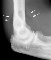
This plain lateral radiograph of the right elbow reveals the classic elbow fat pad sign.
This is an invaluable soft tissue finding in cases of intra-articular injury of the elbow.
Fat is normally present within the joint capsule of the elbow, but outside the synovium.
Typically "hidden" in the concavity of the olecranon and coronoid fossae, the fat is usually not
visible on the lateral radiograph.
However, intra-articular fluid causes distension of the synovium and forces the fat out of the
fossa, producing triangular radiolucent shadows anterior and posterior
to the distal end of the humerus.
Present in a patient with a history of acute trauma to the elbow, the fat pad sign indicates the presence of an intra-articular hemorrhage, which in turn is often associated with an intra-articular skeletal injury (usually the radial head in an adult).
Literature
Siegel MJ: Elbow fat pads with new signs and extended differential diagnosis.
Radiology 1977 Sep;124(3):659-65.
Although fat pad signs are classically associated with
fracture, there are many nontraumatic disease processes which lead to positive fat pad signs. Any intra articular fluid or tissue accumulation
may result in an abnormal posterior fat pad. A "paradoxical" positive
posterior fat pad sign may occur in some instances of extra articular
disease. An extended differential diagnosis is presented.
| [Start] | [Main Elbow] | [A System of Orthopaedic Medicine] |
Copyright © 2021 DR. L. OMBREGT
All Rights
Reserved
The author can not be held responsible for any damage caused by the use of any information
provided.